Frequently asked questions
What are the common symptoms?
What are the common symptoms?
The most common symptoms are difficulty in swallowing, regurgitation of food and choking during eating. People sometimes wake up at night with coughing, due to pooling of food within the diverticulum which can cause frequent regurgitation and can lead to aspiration pneumonia. This occurs when food, saliva, liquids or vomit is breathed into the lungs or airways leading to the lungs.
Zenker’s diverticulum significantly affects people’s quality of life.
Who performs this treatment?
Who performs this treatment?
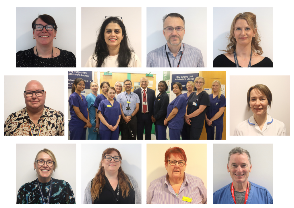
Prof Sauid Ishaq introduced a procedure to treat the condition in the UK in 2013 and we take referrals nationwide. We have won prizes when we have presented our data in national meetings. Patients only have to be in hospital for the day to have the procedure and we are helped by the of anaesthetic team – Dr Andrea Gait and Dr Andy Downs.
Our nursing team is highly skilled in helping with this novel procedure. Team include Hannah Peverell, Jodie Buckley, Jill Harper, Tracey Young, Kay Boyce, and is led by Tracey Ellinor. Dr Adrian Hall from radiology and Linzie Priestnall from SALT team provide support in radiological assessment of the pouch.
What are the treatment options?
What are the treatment options?
Conventional surgical Treatment option
Traditionally ENT colleagues treat ZD with stapling operation that is considered a good treatment but requires general anaesthesia. As metal weerda diverticuloscope is used to access the pouch- placement requires neck extension- that can be challenging in patient with arthritis of neck. Rarely placement can cause broken teeth.
How our flexible endoscopy treatment is different for surgical stapling
Many patient with this condition are elderly with significant health problem hence not fit have general anaesthesia. In addition many patients pouch size is small particularly if patient had treatment before: in this setting conventional treatment if quite challenging with high failure rate. Using Flexible endoscopic approach in such patients is now considered the first line treatment.
Flexible Endoscopic procedure to treat pharyngeal pouch (Zenker’s diverticulum)
In the last 20 years, Zenker’s diverticulum has been treated with a flexible endoscopy. (This is known medically as an endoscopic Zenker’s diverticulectomy.) A flexible telescope is passed over the tongue and down the throat and can be used to identify the pharyngeal pouch (sac) in the back of throat at the level of entrance point into the gullet (food pipe). The bridge of muscle that leads to this sac is identified and is cut with a very small sophisticated knife. The base of this cut muscle part is clipped with a metal clip to prevent any perforation (hole in the linings of the sac). These clips fall out in few days in the stomach and passed in stool.
How effective is Flexible endoscopic treatment zenker diverticulum.
How effective is Flexible endoscopic treatment zenker diverticulum.
This treatment has proved to be effective in over 85 per cent of cases. It is associated with a shorter hospital stay and reduces patient’s discomfort after the procedure. Patients are also able to starting eating again 24 hours after this endoscopic treatment. Therefore, the overall benefit of treating Zenker’s diverticulum with a flexible telescope is very high. One shot of antibiotics is sometimes given in some patients to prevent any infection. 15% of the patient may develop recurrent symptom, again treated in the same fashion with excellent success rate.
What preparation is required?
What preparation is required?
This procedure is carried out with deep sedation with propofol. Patients are required to not eat solids for 24 hours (only liquids) and be nil by mouth (not able to eat) for at least 12 hours before the procedure. Deep sedation is given by expert anaesthetists. Small number of selected patient may require General anaesthesia.
What happens after the procedure?
What happens after the procedure?
Most patients can go home on the same day but a small number will need to be kept overnight for observation. These may include elderly patients, or those with a significant health problem or who have travelled from outside the region)
Are there any risks involving having an endoscopic Zenker’s diverticulectomy?
Are there any risks involving having an endoscopic Zenker’s diverticulectomy?
This procedure is considered safe; however, all procedures carry some risks. The most serious complication is perforation (a hole) at the site of the cut. If this is identified during the procedure, a clip is placed to secure it.
In a published large series of patients (over 500) treated with this procedure, in up to five per cent of the patients, air leaked through the wall of the gullet. If this happens, you will need to stay in hospital and not eat or drink anything until this has healed. During this time, you will be fed with either a small tube through your nose into your stomach or through a drip into your vein in your arm.
You may require antibiotics to prevent infections. As in this procedure we have to cut the muscle, there is a two to three per cent risk of bleeding that can be secured with a clip during the procedure.
What if I have already had surgical treatment of my pouch and symptoms come back – can this be treated?
What if I have already had surgical treatment of my pouch and symptoms come back – can this be treated?
Many of the patients having this procedure are those who have had a recurrence of symptoms after previous surgery.
What if I am on medication to thin my blood?
What if I am on medication to thin my blood?
It is very important, if you are on blood thinning medication, to inform the doctor during consultation as we have to monitor, or in many cases temporarily stop, this medication.
What if I have pacemaker?
What if I have pacemaker?
This usually does not cause any problems with having the treatment, but it is important to inform the treating team.
Research in Zenker diverticulum
Research in Zenker diverticulum
As we are one of the leading centres in world to offer this treatment, we are heavily involved in research and clinical trials to collect data to make this procedure safe and as effective as it can be. Prof Ishaq had led European guideline on endoscopic treatment of Zenker. Please see in our publication section. We may ask for your consent and help in participating in on-going studies. However, this is voluntary and it is totally up to you to enrol or opt out. This will not affect your treatment.
Visiting teams to our centre
Visiting teams to our centre
We train and support experts from other centres to learn this technique, and from time to time, we have experienced endoscopists visit us to learn this procedure. We aim to train at least 6 centres in United Kingdom so patient won’t have to travel far to have this procedure done. These endoscopists may accompany the medical team treating you and you may be asked if you are willing to take part in a teaching/training session that is attended by them. This in any way doesn’t affect the quality of procedure. As a patient, you have an important part in the teaching work of the hospital, but if you do not want students to be involved in your care, please tell the lead nurse and/or doctor when you arrive at the hospital.
Contact details
Contact details
To ask about the procedure call 01384 456111 ext. 2390 (8.00am to 6.00pm, Monday to Friday)
If you have had the procedure and have any concerns call 01384 456111 ext. 2731 (8.00am to 6.00pm, Monday to Friday)
For referrals and general enquiries please email jody.smart@nhs.net
More information (published work from our centre)
More information (published work from our centre)
Ishaq S, Sultan H, Siau K, Kuwai T, Mulder CJ, Neumann H.
Dig Endosc. 2018 Feb 9. doi: 10.1111/den.13035.
Flexible endoscopic treatment for Zenker‘s diverticulum: a systematic review and meta-analysis.
Ishaq S, Hassan C, Antonello A, Tanner K, Bellisario C, Battaglia G, Anderloni A, Correale L, Sharma P, Baron TH, Repici A. Gastrointest Endosc. 2016 Jun;83(6):1076-1089.e5. doi:
Antonello A, Ishaq S, Zanatta L, Cesarotto M, Costantini M, Battaglia G.
Surg Endosc. 20
16 Jun;30(6):2351-7. doi: 10.1007/s00464-015-4482-5
Battaglia G, Antonello A, Realdon S, Cesarotto M, Zanatta L, Ishaq S. Dig Endosc. 2015 Nov;27(7):728-33. doi: 10.1111/den.12490.
Team members
Team members